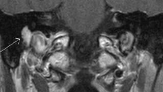
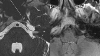
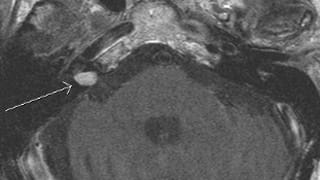
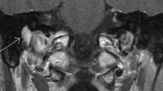
MRI of the Internal Auditory Canals with and without Intravenous Contrast
Study Description
High-resolution MR imaging of the internal auditory canals and petrous temporal bones, as well as MR imaging of the entire brain, with and without intravenous contrast.

Patient Selection and Indications
- Sensorineural hearing loss
- Central origin vertigo
- Facial neuropathy
- Retrotympanic mass
- Postoperative evaluation (for cholesteatoma, schwannoma, etc.)
- Tinnitus (but CT angiogram may be performed for pulsatile tinnitus as well)
- Complicated Bell’s palsy

Patient Preparation
General contraindications to MRI include certain metallic implants and stimulator devices (most cardiac pacemakers, cochlear implants, neurostimulators, aneurysm clips, etc.), as well as certain metallic foreign bodies, such as within the orbits. The MRI technologists screen all patients prior to imaging.
MRI intravenous contrast may be contraindicated in some patients, such as in patients with renal failure on dialysis. The technologists will screen for contraindications to MR intravenous contrast as well.
Claustrophobic patients may require an anxiolytic; severely claustrophobic patients may not be able to tolerate the examination.

Reporting and Outcomes
Thorough evaluation of the brain stem, 7th and 8th cranial nerves, membranous labyrinths, and temporal bones.
Evaluate for potential:
- Benign or malignant masses (vestibulocochlear schwannoma, facial schwannoma, facial nerve hemangioma, meningioma, metastatic disease, brain stem glioma, perineural spread of parotid malignancy, arachnoid cyst, epidermoid cyst, cholesteatoma, cholesterol granuloma, middle ear adenoma, paraganglioma, endolymphatic sac tumor, neurofibromatosis type II)
- Vascular lesions (vascular loop, aneurysm, vascular malformation)
- Infectious or inflammatory lesions (meningitis, Ramsay-Hunt, apical petrositis, labyrnthitis, abscess, neurosarcoidosis)

