MR Defecography
Study Description
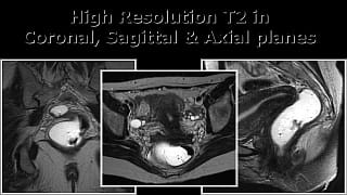
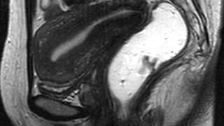
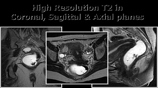
MR Defecography is a procedure performed to evaluate pelvic floor anatomy and function. Routine images of the pelvis are initially obtained and allow evaluation of pelvic organs and musculature. Subsequently, dynamic images are acquired during defecation which enables the radiologist to evaluate organ motion caused by increased pelvic pressure.
With this single examination, information is obtained regarding the anterior compartment (bladder and urethra), central compartment (vagina and uterus) and the posterior compartment (rectum and anus). Most cases of pelvic floor dysfunction involve more than one compartment, and incomplete repair is a common cause of recurrent symptoms.
Unlike traditional evacuation proctography, MR Defecography does not require catheterization or placement of radiodense material in the vagina. A small amount of gel is placed into the rectum just prior to the scan. The patient defecates into an adult diaper while on the MR table, and a drape is used to provide privacy during this portion of the study.
Another advantage of this exam over evacuation procotography is the ability to evaluate incidental pathology of the uterus, ovaries and colon. Furthermore, no ionizing radiation is used during the examination.
Patient Selection and Indications
Common patient symptoms which may be assessed include:
- Urinary retention or incontinence
- Fecal incontinence or obstructed defecation
- Dysfunctional defecation
- Masses found on physical exam which are suspected to be caused by pelvic organ prolapse

Patient Preparation
No special preparations are needed for the examination, and the patient does not have to be NPO.
Standard MR precautions apply for this procedure.
No ionizing radiation is used during this examination.
Prior to the examination, a small amount of gel is placed into the rectum. The patient is imaged in the supine position, and the total examination time is typically around 30 minutes.

Reporting and Outcomes
Pelvic floor anatomy is reviewed in multiples planes to detect injuries to the pelvic floor musculature, which can be frequently seen in patients following vaginal delivery. Pelvic organs such as the uterus and ovaries are evaluated for pathology.
A set of images is obtained during rest, Kegel maneuver and Valsava maneuver. The anal canal/pubococcygeal angle is measured on all three sets of images to determine if pelvic floor motion is appropriate.
The last phase of the exam consists of the patient evacuating gel from the rectum while being imaged. This provides the best visualization of pelvic floor weakness, particularly the development of organ protrusion or intussusception. Organ descent is measured with reference to the pubococcygeal line, and qualitative assessment of rectal evacuation is given.
All of these exams are reviewed by radiologists with subspecialty training in advanced body imaging. Any urgent finding will be communicated by phone to the referring physician.

