fMRI
Study Description
- No contrast required
- Non-invasive MRI exam
- No radiation
- Blood oxygen level dependent functional MRI sequence
- Various imaging paradigms
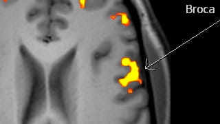
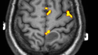
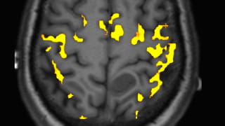
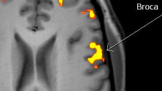
- A MR sequence which measures ↑ blood flow to areas of the brain being used/activated ultimately providing an image/map of the area of the brain performing the task.

Patient Selection and Indications
Pre-operative evaluation for patients with intra-cranial lesion or neoplasm prior to resection or biopsy, or to help determine if lesion can be safely resected.
- Determine tumor relationship to motor, language (Wernicke and Broca), visual or auditory centers in the brain to help guide biopsy or resection approach and decrease procedural morbidity.
- Determine hemispheric dominance for language (replaces WADA test) in right and left handed patients to help decide if biopsy or resection can be performed, guide safer resection and help aid in preoperative risk of procedure explanation to the patient

Patient Preparation
- The exam is performed at the Wellstar Kennestone Hospital 3T MRI unit.
- The patient needs to be lucid enough to perform the fMRI paradigms. This involves following directions regarding finger tapping or foot movement (for motor), sentence reading or word/verb generation sentence completion (for language), procession visual stimuli (vision), or listening to sounds (auditory).
- Exam is approximately 30 minutes and patient needs to be able to lie still in the MRI bore.
- Patient should not be claustrophobic.
- Patient cannot have implantable medical devices (such as pacemaker etc.) or other metal in body (i.e. eyes) which would be a contraindication to MR.

Reporting and Outcomes
- Images are post-processed at the 3T MR suite workstation in conjunction with a neuroradiologist.
- fMRI images are fused onto a volumetric high resolution TI sequence of the brain.
- The study is reviewed, discussed with the neurosurgeon and dictated by a neuroradiologist.

