CT Urography
Study Description
CTU is a CT scan of the abdomen and pelvis with and without contrast that utilizes a special injection technique to evaluate both the renal parenchyma and urothelial systems at the same time.
Traditional evaluation of symptoms such as hematuria required multiple radiologic exams such as renal ultrasonography, CT of the abdomen and pelvis with and without contrast, and intravenous pyelography (IVP). CT Urography has replaced IVPs at most centers throughout the US, due to its ability to evaluate for the presence of stone disease, renal masses and upper tract disease in one comprehensive, fast, well tolerated examination. In addition, CTU also provides the same evaluation of the abdominal and pelvic organs as a routine CT scan, and superior mass evaluation when compared to ultrasound.
CTU produces a radiation dose similar to a routine with and without contrast CT of the abdomen and pelvis. Patients receive an initial non-contrast scan followed by a split bolus injection of intravenous contrast. The bolus is split to allow optimal parenchymal enhancement of the kidney (nephrographic phase) at the same time that the patient is excreting the first contrast bolus in the urine (excretory phase), allowing for evaluation of the collecting system/ureters/bladder during the same scan.

Patient Selection and Indications
Evaluation of both the renal parenchyma and urothelial system for:
- Unexplained hematuria
- Genitourinary anomaly evaluation
- Procedural complications
- Traumatic injury

Patient Preparation
No oral contrast is needed.
- 900 cc’s H2O Orally 1/2 hour before the scan while at the performing facility.
- This exam requires IV contrast
- Patient NPO for 4 hours
- 5mg of Lasix IV
Standard precautions with regard to IV iodinated contrast are necessary.

Reporting and Outcomes
The non-contrast portion of the scan is used to evaluate for renal and ureteral calculi, as well as providing baseline density measurements for masses.
The contrast portion of the exam is used to evaluate all abdominal and pelvic organs, with special emphasis on renal parenchyma and masses. Renal vascular anatomy can also be evaluated, although not with the same sensitivity as a CT angiogram.
During the contrast portion, the urothelial system is evaluated for masses and congenital abnormalities.
While the examination surpasses that of conventional imaging studies, cystoscopy still provides superior evaluation of the bladder.
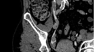
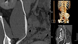
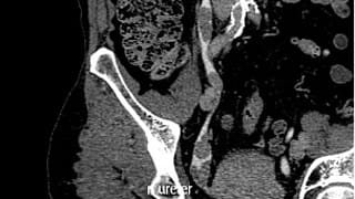
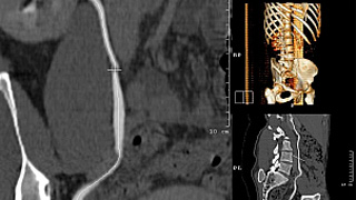
All CT Urograms are post processed by the Q3D Laboratory, with true coronal views of the kidneys, rotational images of the ureters and 3D views of the genitourinary system.
All studies will be interpreted by radiologists with subspecialty training in advanced body imaging. Urgent results will be communicated at the time of dictation.

